1 EYE, EAR, NOSE, AND THROAT (EENT) SURGERY
Section I. Eye Surgery
Section II. Ear Surgery
Section III. Nose Surgery
Section IV. Throat, Tongue, and Neck Surgery
2 PROCEDURES IN GYNECOLOGICAL AND OBSTETRICAL SURGERY
Section I. Anatomy of the Female Reproductive System
Section II. Vaginal Surgery
Section III. Abdominal Gynecological and Obstetrical
Surgery
3 PROCEDURES IN GENITOURINARY SURGERY
Section I. Anatomy and Physiology of the
Genitourinary Organs
Section II. General Considerations in Genitourinary
Surgery
Section III. Operations on the Kidney, Ureter, and
Adrenal Glands
Section IV. Operations on the Bladder and Prostate
Section V. Operations on the Scrotum, Penis, and
Urethra
Section I. EYE SURGERY
1-1. INTRODUCTION
a. General.
The anatomy, physiology, and the location of the eye make surgery upon
the eye a highly specialized field of surgery. Therefore, procedures
done by the specialist when assisting with eye surgery differ from
procedures used for other surgical specialties. However, the
principles of asepsis and safe, skillful care apply as in all other
surgery. The ensuing text presents a discussion of the necessary
considerations that are applicable in the majority of cases in this
specialty.
b. Special Care of Instruments.
The specialist is to use exacting care
when working with instruments for eye, ear, nose, and throat surgery
because most of these instruments are delicate. Sharp surfaces of
these instruments must be preserved to ensure the success of the
operative procedure. The specialist is to follow local policy in the
care and handling of these instruments.
c. Anatomy and Physiology of the Eye.
The eye is also referred to as the
eyeball or globe. In the adult, it is slightly less than one inch in
its longest diameter. See figure 1-1 for parts of the eye.
(1) The lids and anterior surface of the eye, except
for the center, are covered by the conjunctiva.
(2) The cornea forms the anterior center of the eye
and transmits and refracts light. Behind it, the anterior chamber
contains the iris (which gives eye color and forms the pupil) and
the aqueous humor.
(3) The lens focuses light on the retina allowing
for near and far vision.
(4) The posterior chamber contains the jelly-like
vitreous humor, which helps give rigidity to the eye.
(5) The retina receives light and converts it to
impulses to the brain via the optic nerve.
(6) The main body of the eye is made of three layers called
tunics. The external tunic includes the sclera (the white part of
the eye) and clear cornea. The middle tunic includes the choroid,
the ciliary body, and the iris. The iris is the colored part that
changes the aperture size over the eye lens. The internal tunic is
sometimes called the nervous covering, but is usually referred to as
the retina. The retina is a thin network of nerve cells and fibers
that receives the images of objects the eye is seeing.
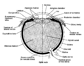
Figure 1-1. Parts of the eye.
1-2. SPECIAL PREPARATION OF THE OPERATING ROOM
a.
Instruments. All instruments used for eye surgery are made
for this purpose, and are unlike those for surgical procedures in
other areas of the body. Preferences for instruments vary so widely
among eye surgeons that it may be necessary to list all instruments
used for each operation by each different surgeon. Therefore, the
surgeon's card must be carefully checked when selecting instruments
for an eye operation.
b. Sponges.
Gauze sponges are considered much too rough for use on an
eyeball. Instead, dampened cotton applicators are used. Special
cellulose sponges, specifically designed and prepackaged sterile by
manufacturers for eye surgery, are also available.
c. Magnifying Glasses.
The surgeon may wish to use special magnifying glasses during
the procedure; therefore, these must be cleansed and ready for use.
d. Lighting.
Illumination for eye surgery may be furnished by a number of methods.
(1) One method is the use of the standard overhead
light. The circulator may be responsible for adjusting the light
during surgery. If this need occurs, he should pay particular
attention to not contaminating the sterile field and scrubbed
personnel.
(2) A second source is the use of an electric head
lamp. This lamp is strapped to the surgeon's head and is used in the
same manner as a coal miner's helmet. The surgeon may redirect the
light during surgery.
(3) The third method is the use of the operating
microscope. This is a device used to magnify the site of surgery and
enable the surgeon to do very delicate work with excellent
illumination. This device is draped with sterile material before the
procedure is started, and the surgeon may make any adjustments. The
microscope is being used more and more for eye and other delicate
surgery.
e. Medications.
As many as 5 or 6 solutions may be kept within the sterile field for
use during eye procedures; examples of these are saline (for dampening
the eyeball), local anesthetic agents, and epinephrine. If these are
not prepackaged and sterilized in individually labeled doses, the
specialist should label medicine glasses to show the name and the
strength of each solution. During preparation for an operation, the
circulator should pour the solutions needed into the medicine glasses,
making sure that the solution he is pouring matches the label on the
glass. Great care should be taken to assure that ophthalmic solutions
of the desired drugs are used.
f. Sterile
Setup. If both of the patient's eyes are to be operated on
for correction of defects requiring muscle surgery or other
extraocular procedures, only one Mayo table needs to be up. However,
if intraocular surgery is to be performed on both eyes, the specialist
sets up two tables--one for each eye. When the procedure on the first
eye is completed, the surgeon and specialist change only their gloves
in preparation for the second eye.
NOTE: A large percentage of intraocular
surgery does not require double setups. Advancement in techniques and
equipment makes the practice ineffective and costly.
From Special
Surgical Procedures II