INTRODUCTION
1 THE REPRODUCTIVE SYSTEMS
Section I. The Female Reproductive System
Section II. The Male Reproductive System
Section III. Events of Pregnancy
Exercises
2 NORMAL AND EMERGENCY CHILDBIRTH
Section I. General Information
Section II. Complications of Pregnancy
Section III. Management of Mother and Newborn During
Normal Delivery in an Emergency Setting
Section IV. Abnormal Deliveries
Section V. Complications of Labor and Delivery
Exercises
3 PEDIATRIC EMERGENCIES
Section I. Differences Between a Child's Body and an
Adult's Body
Section II. Patient Assessment
Section III. Special Considerations of the Ill or
Injured Child
Section IV. Pediatric Emergencies
Section V. Trauma in Children
Exercises
4 CHILD ABUSE
Exercises
----------------------------------------------
LESSON 1
THE REPRODUCTIVE SYSTEMS
Section I. THE FEMALE REPRODUCTIVE SYSTEM
1-1. INTRODUCTION
a. Reproduction Defined. The mechanism by which
life is maintained is reproduction. Reproduction can be defined as the
process by which a single cell duplicates its genetic material, thus
allowing an organism to grow and repair itself.
Reproduction, therefore, maintains the life of a
member of a species. Additionally, reproduction is the process by
which genetic material is passed from generation to generation.
b. Major Types of Reproduction. There are two
major types of reproduction: asexual and sexual. Only one parent is
involved in asexual reproduction. The parent cell may divide and
become two new cells, or the new organism may arise from a part of the
parent cell. In the case of humans, sexual reproduction takes place.
This requires the participation of two parents. Each parent produces
special reproductive cells called sex cells or gametes. In this sense,
reproduction maintains the continuation of the species. If a species
loses its reproductive capability, the species no longer survives. It
becomes extinct.
c. Female Reproductive System Functions. The
female reproductive system has specialized organs to carry out its
three important functions. These functions are the production of egg
cells, the disintegration of nonfertilized egg cells, and the
protection of the developing embryo.
1-2. EXTERNAL GENITALIA
The vulva and its parts make up the external
genitalia. The word vulva is a term that has been designated to stand
for the external genitalia of the female.
a. Mons Pubis. The elevated, fatty tissue
covered with coarse pubic hair which lies over the symphysis pubis is
the mons pubis. Pubic hair appears at puberty. The function of the
mons pubis is to protect the pelvic bone.
b. Labia Majora. The labia majora are large,
longitudinal folds of skin and fatty tissue which extend back from the
mons pubis to the anus. The outer surfaces are covered with hair. The
inner surfaces are smooth and moist. The corresponding structure in
the male is the scrotum. The function of these folds is to protect the
entrance to the vagina.
c. Labia Minora. The labia minora are two folds
of skin lying within the labia majora and also enclosing the
vestibule. In front, each labium minus (minus = singular of minora)
divides into two folds. The fold above the clitoris is called the
prepuce of the clitoris. The fold below is the frenulum. No pubic hair
is on these structures.
d. Clitoris. The clitoris is a small projection
of sensitive, erectile tissue which corresponds to the male penis. The
female urethra, however, does not pass through the clitoris. As in the
male penis, the clitoris is covered by prepuce.
e. Urinary Meatus. The urinary meatus is the
small opening of the urethra which is located between the clitoris and
the vagina.
f. Vaginal Orifice. This is the opening to the
vagina from the outside.
g. Bartholin's Glands. These are bean-shaped
glands located on each side of the vaginal orifice. They provide
lubrication of the vagina.
h. Perineum. The perineum is the area between
the vaginal orifice (opening) and the anus.
1-3. INTERNAL GENITALIA
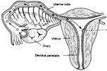
a. Uterus or Womb.
(1) Description/information. The uterus is a hollow,
muscular, pear-shaped organ. It is located in the pelvic cavity
between the urinary bladder and the rectum. During a woman's
child-bearing years, the uterus is about 7.5 centimeters long, 5
centimeters wide, and 2.5 centimeters thick. The uterus has three
anatomical divisions: the fundus, the body, and the cervix. The fundus
is the upper, convex part of the uterus. This part is just above the
entrance to the uterine tubes. The body is the central portion of the
uterus, and the cervix is the lower, neck-like part of the uterus.
(2) Walls. The walls of the uterus are made up of
three layers: the endometrium, the myometrium, and the parietal
peritoneum. The endometrium, the inner layer, attaches itself to the
myometrium layer and lines the uterus. This layer is sloughed off
during menstruation or post- delivery. The middle layer, which is
composed of smooth muscle, is the myometrium. This layer is made up of
longitudinal, circular, and spiral muscular fiber which interlaces.
The myometrium is thickest in the fundus and thinnest in the cervix.
During childbirth, this muscle layer is capable of the very powerful
contractions necessary for a normal birth. The third layer, the
parietal peritoneum, is the outer layer which is a serous membrane.
This outer layer of uterine wall is incomplete, covering only part of
the uterine body and none of the uterine cervix.
(3) Functions. The uterus has three major functions
which occur during these events: pregnancy, labor, and menstruation.
During pregnancy, the uterus holds the fertilized ovum. The ovum is
deposited in the uterus where it grows and develops through the embryo
and fetal stages. During the birth process, the uterus produces
powerful contractions to expel the mature infant. And, finally, during
a female's menstrual phase, the inside lining of the uterus detaches
and sloughs off, the uterus expelling its fluid contents.
b. Uterine Tubes, Fallopian Tubes, or Oviducts.
(1) Description/information. These tubes are known by
all three names listed above. The name commonly used is fallopian
tubes. These two tubes extend from the ovaries to the uterus. An ovum
discharged from an ovary passes through one of these tubes to the
uterus. Each tube is about 10 centimeters long (4 inches). The tube is
located between the folds of the broad ligaments of the uterus. The
tubes are attached to the uterus at one end but not attached to the
ovaries at the other end. At the ovary end, the tubes are open,
funnel-shaped, and close to the ovary. The funnelshaped ends of the
tubes are called the infundibulum, and the fringe or finger-like
processes at the tube ends are called fimbriae.
(2) Functions. The uterine tubes are ducts for the
ovaries although the tubes are not attached to the ovaries.
Additionally, the tubes are the site of fertilization. Fertilization
normally takes place in the outer one-third of the tube.
c. Ovaries.
(1) Description/information. The ovaries are two
almond-shaped glands. They are located on either side of the uterus,
below and behind the uterine tubes. The ovaries are detached from the
uterine tubes and held in position by a series of ligaments. During
the second phase (preovulatory phase) of the menstrual cycle, one of
the 20 to 25 primary follicles developed during the menstrual phase
matures into a Graafian follicle, a follicle ready for ovulation.
During the maturation process, this follicle increases its estrogen
production. The rupture of the Graafian follicle with the release of
the ovum is the beginning of ovulation.
(2) Functions. One function of the ovaries is to
produce ova (female reproductive cells capable of developing, after
fertilization, into new individuals). Also, the ovaries discharge ova
(ovulation) and secrete the female sex hormones progesterone,
estrogen, and relaxin. The ovaries in the female correspond to the
testes in the male reproductive system.
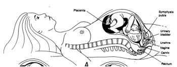
d. Vagina.
(1) Description/information. The vagina is a muscular,
tubular organ lined with mucous membrane. This organ is about 10
centimeters (4 inches) long and extends from the hymen to the cervix.
The vagina extends upward and backward between the rectum and the
bladder and is attached to the uterus.
(2) Structure. The lining of the vagina is made up of
smooth muscle which is longitudinally and circularly arranged in many
folds called rugae. The folds of the lining permit the organ to expand
when necessary. The hymen is the fold of mucous membrane at the
orifice (opening) of the vagina.
(3) Functions. The vagina serves as a passageway for
menstrual flow, receives seminal fluid from the male, and serves as
the lower part of the birth canal.
From
Obstetrics/Pediatrics