|
CORRESPONDENCE
COURSE
ACADEMY OF HEALTH SCIENCES, UNITED STATES ARMY
SUBCOURSE MD0575 EDITION 100
INTEGUMENTARY SYSTEM
The skin is not just a simple thin covering which keeps the body
together. The skin is a complex combination of tissues that perform
functions necessary for human survival.
Our skin helps maintain body temperature, receives stimuli from the
environment, and stores chemical compounds. Consider the human
predicament if the skin were not waterproof. Every time it rained,
each human would absorb water like a sponge.
The skin also acts as a protective covering keeping underlying tissues
from bacterial invasion and harmful light rays and from drying out.
As a Medical NCO, it is important for you to understand the complex
functions of the skin.
----------------------
Length: 97 Pages
Estimated Hours to Complete: 8
Format: PDF file
Size: 1.2 MB
----------------------------
Anyone may take this course. However, to receive credit hours, you
must be officially enrolled and complete an examination furnished by
the Nonresident Instruction Branch at Fort Sam Houston, Texas.
Enrollment is normally limited to Department of Defense personnel.
Others may apply for enrollment, but acceptance is not guaranteed.
The Integumentary System
Distance Learning
Course
97 Pages
Est. 8 Hours
1.2 MB pdf file
Download Now |
|
TABLE OF CONTENTS
INTRODUCTION
1 ANATOMY AND PHYSIOLOGY OF THE INTEGUMENTARY SYSTEM
Exercises
2 PHYSICAL ASSESSMENT OF THE INTEGUMENTARY SYSTEM
Exercises
3 PRIMARY AND SECONDARY SKIN LESIONS
Exercises
4 COMMON SKIN DISEASES
Exercises
5 DERMATOLOGICAL DRUGS
Exercises
---------------------------------------
LESSON 1
ANATOMY AND PHYSIOLOGY OF THE INTEGUMENTARY SYSTEM
1-1. INTRODUCTION
a. The integumentary system, consisting of the skin
and its derivatives, is the largest and one of the most complex
systems of the body. The surface area of the skin covers about 1.8
square meters (19.4 square feet) of the body of the average male
adult. The skin weighs about six pounds and receives roughly one-third
of all blood circulating through the body. It is difficult to think of
the skin as a system, but it is a complex of organs (sweat glands, oil
glands, and so forth). It is elastic, regenerates, and functions in
protection, thermoregulation, and sensation.
b. The protection, sensations, secretions, and the
other functions which the integument gives to the rest of the body are
essential for life. Changes in the normal appearance of the skin often
indicate abnormalities or disease of body function. As a medical
non-commissioned officer (NCO), you need to recognize changes in skin
appearance that your treatment might affect. A basic knowledge of the
normal anatomy and physiology of the integumentary system is essential
to your job.
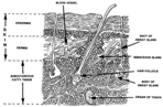
1-2. LAYERS OF SKIN
a. General Information.
Skin consists of three distinct layers: the
epidermis, the dermis, and the subcutaneous layer
(figure 1-1). The top layer, the epidermis, is attached to the second
layer, the dermis. The dermis is thick, connective tissue. Individuals
with thick skin have a relatively thick epidermis. Persons with thin
skin have a thin epidermis. The subcutaneous layer, the third layer of
skin, is located beneath the dermis and consists of areolar (minute
spaces in tissue) and adipose (fat) tissues. The first skin layer is
fixed to the second skin layer as though the two were glued together.
The second and third skin layers are attached in a different way.
Fibers from the second layer (the dermis) extend down into the third
layer (subcutaneous), anchoring the two layers together. The third
layer is firmly attached to underlying tissues and organs of the body.
b. Epidermis.
(1) The epidermis is composed
of stratified, squamous (scale-like), epithelial cells which are
organized in four or five layers. The number of cell layers depends on
the location of the skin on the body. The epidermis has five layers on
the palms of the hands and the soles of the feet because those areas
have more wear and tear. Skin on other parts of the body has four
layers of epidermis because there is less exposure to frictions.
(2) These are the layers of the epidermis
(figure 1-2) from the deepest to the most superficial.
(a) Stratum basal. Cells
continually multiply and push upward toward the surface.
(b) Stratum spinosum. Eight to ten rows of
polyhedral (many sided) cells which fit closely together make up this
layer of epidermis. New cells germinate in this layer.
(c) Stratum granulosum. Three
to five rows of flattened cells containing keratohyalin, a substance
that will finally become keratin, make up this layer of epidermis. The
nuclei of cells are in various stages of degeneration--breaking down
and dying.
(d) Stratum lucidum. This
layer is thicker on the palms and soles. The layer consists of several
rows of clear, flat, dead cells that contain droplets of a clear
substance called eleidin. Eleidin eventually becomes keratin.
(e) Stratum corneum.
Twenty-five to thirty rows of flat, dead cells that are completely
filled with keratin make up this layer. These cells are shed and
replaced continuously so that roughly every twenty-eight days, this
layer is new. It is this layer with its water-proofing protein keratin
which keeps the body from soaking up water like a sponge. These
keratin-filled, dead cells serve as a barrier against light and heat
waves, bacteria, and many chemicals.
c. Dermis.
(1)
Composition. The second layer of skin, the dermis or corium, is
sometimes called the true skin. It holds the epidermis in place by
connective tissue and elastic fiber. The dermis is very thick on the
palms of the hands and the soles of the feet but very thin on the
eyelids, penis, and scrotum. The dermis contains the following:
numerous blood vessels, nerves, lymph vessels, hair follicles, sweat
glands, and sensory receptors.
(2)
Dermis layers.
(a) Papillary layer. This
upper one-fifth of the dermis has small, finger-like projections
called dermal papillae. These projections reach into the concavities
between ridges in the deep surface of the epidermis. This region or
layer consists of loose connective tissue containing fine elastic
fibers.
(b) Reticular layer. This
layer makes up the rest of the dermis. The reticular layer consists of
dense, irregularly arranged connective tissue which has interlacing
bundles of collagenous and coarse fibers. Between the fibers are
adipose (fat) tissue, hair follicles, nerves, oil glands, and the
ducts of sweat glands. The collagenous and elastic fibers together
give the skin strength, extensibility, and elasticity.
NOTE: Extensibility is the
ability to stretch. Elasticity is the ability to return to original
shape after extension or contraction.) The skin stretches during
pregnancy, obesity, or edema. Elasticity allows the skin to contract
after such stretching. If the skin has been stretched severely, small
tears may occur. Initially, the tears are red; they lose the redness
but remain visible as silvery white streaks called striae.
d. Subcutaneous-Adipose.
This layer is composed of loose connective tissue combined
with adipose (fatty) tissue. The subcutaneous layer of skin has
several important functions:
(1)
Storehouse for water and particularly for fat. Much of the fat in an
overweight person is in this layer.
(2)
Layer of insulation protecting the body from heat loss.
(3)
Pads the body giving the body form and shape and cushioning and
protecting the body from blows.
(4)
Provides a pathway for nerves and blood vessels.
From The
Integumentary System |
